
Revision History
The ACR Breast Ultrasound Accreditation Program is facility-based. It is comprised of two modules: the Breast Ultrasound module, which includes Cyst and Solid submissions, and the Breast Ultrasound Biopsy module, which includes Core Needle Biopsy (CNB) and/or Fine Needle Aspiration Cytology (FNAC) submissions. Facilities must apply for accreditation for all services provided. For example, facilities that do not perform CNB or FNAC procedures are only required to apply for the Breast Ultrasound module. If the facility performs CNBs or FNACs of solid masses (excluding dermal lesions, cyst aspirations, and intramammary and axillary lymph nodes), then the Breast Ultrasound Biopsy module must also be applied for with the applicable exams submitted. Only one set of images per type (e.g., simple cyst, solid mass, CNB and/or FNAC) is required, regardless of the number of breast ultrasound units or the number of physicians at the facility.
If it is determined that you need to change exams, the change request must be submitted as a support ticket request before submitting your testing package. There you may select the topic/category of exam change, enter the modality ID number, and specify the exam or module you wish to add, remove, or change. You will be contacted once the change is complete to upload and submit your testing package.
You must log into the ACRedit Plus system to enter and submit data into your online testing package.
During the accreditation review, at least two radiologist reviewers will assess the following aspects of each case:
- Visualization of the cyst or solid mass in the correlating mammogram
- Ability of the facility's radiologist to appropriately triangulate the cyst or solid mass
- Image quality
- Exam identification and labeling
Important Considerations When Selecting Clinical Cases
- Submit examples of your work with the best image quality. If possible, submissions should avoid subtle lesions. The ACR Committee on Breast Ultrasound Accreditation understands that all images obtained during all ultrasound exams or ultrasound-guided breast biopsy procedures may not meet these criteria. Consequently, sufficient time is allowed to select cases that are examples of "best work". ACR reviewers will evaluate your images accordingly.
- Do not submit images that are performed on models or volunteers.
- Do not upload mammography, ultrasound, or pathology reports into the testing package; reports will not be sent to the ACR reviewers. Please note that the ACR will purge any submitted materials containing protected health information (PHI) that are not necessary for the accreditation review process.
- Your supervising physician must review and approve images prior to submitting them to the ACR for review. The supervising physician is ultimately responsible for previewing the images submitted for review.
- All exams submitted must have been performed within six months of the date that the testing package link was released to the facility. No images will be accepted for review that predate this by more than six months.
- All images for each exam must be from the same patient.
- Do not submit implant cases.
- As a general rule, do not submit male patients. If there are exceptional circumstances, please reach out to ACR staff in advance of submission.
- The area being evaluated or biopsied must be visible and easily identified on all submitted images:
- Correlating mammogram - the lesion must be clearly circled (please refer to the requirements below)
- All ultrasound images (please refer to the requirements below)
- Do not anonymize images.
Correlating Mammogram
A full 2-view, 2D and/or 2D synthesized mammogram (CC and MLO/ML/LM) in original DICOM format is required.
- The mammogram may be from an outside facility.
- The mammogram must have been performed within 60 days prior to the ultrasound.
- The required mammogram views (CC and MLO/ML/LM) must be from the same date of service.
- If both the CC and MLO/ML/LM views were not obtained on the same date of service, a different study should be selected for submission.
- Do not submit post-procedure images (e.g., post clip images).
- Include the entire breast with the lesion being evaluated; the lesion should be well-visualized and clearly circled on each image.
- If you do not circle the lesion, the exam will be returned as the reviewers will not be able to determine if the intended lesion was imaged.
- Circling more than one lesion, either with multiple ROIs or with a single ROI that contains multiple lesions, will result in accreditation failure because ACR reviewers may be uncertain which lesion was evaluated or biopsied.
- The ROI must be circled; indicating the ROI with a skin/radiopaque marker, square, or other symbol is not acceptable.
- No other annotations (e.g., measurement calipers) should be inside the circled ROI.
- Additional images (e.g., individual slices from tomosynthesis, XCCL, spot views) are acceptable ONLY in addition to the full 2-view, 2D and/or 2D synthesized CC/MLO/ML/LM mammograms.
- Images must allow the ACR reviewers to determine if the physician can appropriately triangulate the lesion.
Ultrasound Images
- Only submit the requested ultrasound images. Do not submit additional images. This may lead to a delay in the review process as ACR reviewers will only evaluate the images required for accreditation submission.
- Gray scale only images should be submitted.
- Color Doppler images, including color Doppler images with the color box removed, are not acceptable for accreditation review and should not be submitted.
- Harmonic images, as well as compound spatial imaging, are acceptable.
- Split screen images are not acceptable for accreditation review. All ultrasound images must be full screen images.
- You may submit the same case for both the Breast Ultrasound Solid Mass and the Breast Ultrasound Biopsy module Core Needle Biopsy exams or the Breast Ultrasound Solid Mass and the Breast Ultrasound Biopsy module Fine Needle Aspiration Cytology exams. You must submit the images separately in the appropriate modules for each exam.
- All ultrasound images must be taken from the same date of service, including images submitted for Core Needle Biopsy and/or Fine Needle Aspiration Cytology exams.
- Transducers should be operated at a center frequency of at least 12 MHz, and preferably higher. See Equipment: Breast Ultrasound for more information.
- Caliper measurement of the cyst or solid mass should be in its longest dimension; including two caliper measurements is acceptable.
- In alignment with the most recent BI-RADS® guidance, if a solid mass has an echogenic rind, the rind should ideally be included in the measurement.

Breast Ultrasound Module
- Simple Cyst:
- The cyst must meet the BI-RADS® criteria for a simple cyst: a) anechoic, b) circumscribed margin, c) posterior enhancement, and d) round/oval
- For accreditation purposes, do not select a complicated cyst, complex cystic and solid mass, or dermal lesion for submission.
- The simple cyst submitted for accreditation should be over 0.5 cm in the longest dimension.
Simple Cyst Exam Images |
| Full 2-view, 2D and/or 2D synthesized mammogram in original DICOM format with the cyst circled |
| 2 orthogonal ultrasound images (e.g., transverse and longitudinal or radial and antiradial) with no calipers |
| 1 ultrasound image, in either plane, with the appropriate caliper measurement showing the cyst in its longest dimension |
- Solid Mass:
- For accreditation purposes, do not select an intramammary or axillary lymph node, dermal lesion, or complex cystic and solid mass for submission.
Solid Mass Exam Images |
| Full 2-view, 2D and/or 2D synthesized mammogram in original DICOM format with the solid mass circled |
| 2 orthogonal ultrasound images (e.g., transverse and longitudinal or radial and antiradial) with no calipers |
| 1 ultrasound image, in either plane, with the appropriate caliper measurement showing the solid mass in its longest dimension |
Breast Ultrasound Biopsy Module (applies to CNB and FNAC)
- Choose only BI-RADS® Category 4 or 5 cases to submit for accreditation review.
- Images submitted for the breast ultrasound biopsy module should demonstrate that physicians performing these procedures possess the skill necessary for appropriate needle positioning.
- The pre-biopsy and post/post-fire/post-biopsy ultrasound images must be from the same date of service.
- Important labeling requirements for CNB and FNAC - In addition to the labeling requirements for all images included in the Required Exam ID and Labeling table below, please note the following:
- Each biopsy image MUST include the written transducer orientation. Failure to include the written transducer orientation on ALL ultrasound images is cause for failure.
- The oblique orientation is acceptable only for images demonstrating the needle, if appropriate. The orthogonal pre-biopsy images cannot use the oblique label due to the inability to accurately label orthogonal planes from an oblique angle.
- Biopsy images demonstrating the needle must be annotated as Post, Post-Fire, or Post-Biopsy; failure to include Post, Post-Fire, or Post-Biopsy as part of the image labeling is cause for failure.
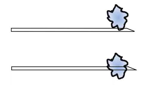
- The position of the needle relative to the solid mass must be easily visualized on all biopsy images. All biopsy images should demonstrate the entire needle (from its base to its tip) in its long axis, approximately parallel to the chest wall.
- For CNB, the post/post-fire/post-biopsy image should demonstrate the needle tip below or through the solid mass. For FNAC, the post/post-fire/post-biopsy image should demonstrate the needle tip in the lesion.
CNB:
FNAC:
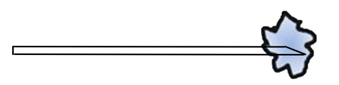
Core Needle Biopsy (CNB):
- For accreditation purposes, do not select an intramammary or axillary lymph node or dermal lesion.
Core Needle Biopsy Exam Images |
| Full 2-view, 2D and/or 2D synthesized mammogram in original DICOM format with the solid mass circled |
| 2 orthogonal pre-biopsy ultrasound images (e.g., transverse and longitudinal or radial and antiradial) with no calipers |
| 1 post/post-fire/post-biopsy ultrasound image demonstrating the needle and mass |
Fine Needle Aspiration Cytology (FNAC):
- Solid masses only - do not select a cyst aspiration, an intramammary or axillary lymph node, or dermal lesion.
Fine Needle Aspiration Cytology Exam Images |
| Full 2-view, 2D and/or 2D synthesized mammogram in original DICOM format with the solid mass circled |
| 2 orthogonal pre-biopsy ultrasound images (e.g., transverse and longitudinal or radial and antiradial) with no calipers |
| 1 post/post-fire/post-biopsy ultrasound image demonstrating the needle and mass |
Exam Identification and Labeling
Images are an important part of the medical record. One of the requirements for clinical images is correct labeling to include patient identification. The ACR understands that as providers, facilities are subject to the Health Insurance Portability and Accountability Act of 1996 (HIPAA) and that is why the ACR executes a HIPAA business associate agreement (BAA) with facilities. This agreement allows the collection of patient information in the performance of ACR accreditation activities which are specifically mentioned in the HIPAA regulations. If the facility has a BAA with ACR, they are covered under HIPAA. If not, contact the ACR to obtain an agreement for signature.
Each image should include the information below:
Required Exam ID and Labeling If any of these items are absent, the case will fail accreditation |
|
| Recommended Exam ID and Labeling |
|
Completing the Testing Package
Log into the ACRedit Plus system and access the testing package. Complete the Patient Outcome Data with information from the prior 12 months or last calendar year, fill out all required clinical data forms, upload a current annual survey for all active units, upload only the required images, and submit the online testing package.
Uploading Images
Submission of compressed DICOM images (e.g., lossy compressed) is not recommended. Compressed images may result in loss of image quality. All submitted images will be scored accordingly.
1. Correlating mammogram
Option 1 (preferred):
- Upload the full 2-view, 2D and/or 2D synthesized mammogram (CC and MLO/ML/LM) in original DICOM format. Each image must include the circled ROI of the lesion being evaluated.
- Additional images (e.g., individual slices from tomosynthesis, XCCL, spot views) may be submitted ONLY in addition to the required full 2-view, 2D and/or 2D synthesized CC/MLO/ML/LM mammogram.
- Please note, secondary capture images and screen shots (e.g., jpeg images) are not original DICOM images.
Option 2: If you are unable to produce mammogram images with the required ROIs in original DICOM format with all required labeling:
- Upload the full 2-view, 2D and/or 2D synthesized mammogram (CC and MLO/ML/LM) in original DICOM format without circled ROIs.
AND
- Upload non-DICOM screen shots of the full 2-view, 2D and/or 2D synthesized mammogram (CC and MLO/ML/LM). Each image must include the circled ROI of the lesion being evaluated.
- Additional images (e.g., individual slices from tomosynthesis, XCCL, spot views) may be submitted ONLY in addition to the required full 2-view, 2D and/or 2D synthesized CC/MLO/ML/LM mammogram.
- Please note, secondary capture images and screen shots (e.g., jpeg images) are not original DICOM images.
2. Ultrasound images:
- DICOM format only
- Only the requested images
- Please do not submit additional images
3. Once uploaded, check all images for adherence to accreditation criteria by clicking "view images" on the "Upload Images" page in the online testing package.
After uploading the required full 2-view, 2D and/or 2D synthesized mammogram images, you may verify that the images are in original DICOM format by using the Ruler tool in the NilRead viewer, which will provide an accurate measurement. Secondary capture images and screenshots (e.g., jpeg images) will not support measurements and do not meet the requirements for original DICOM format.

4. Submit the testing package.
Common Reasons for Image Rejection
- Mammogram images not in original DICOM format
- Providing only tomosynthesis slices and/or spot/compression mammogram images
- Mammogram without the ROI circled on the full 2-view, 2D and/or 2D synthesized CC/MLO/ML/LM views
- Not submitting ultrasound images in two orthogonal planes
- Submitting split screen or color Doppler ultrasound images
Revision History for this Article | ||
Date | Section | Description of Revision(s) |
12-12-19
| All
| Article created; FAQs incorporated; No criteria changes
|
11-15-2022 | Added data sheet attachment | |
| 3-3-2023 | Updated data sheet attachments | |
| 6-8-2023 | Introduction | Removed reference to printing data sheets if submitting by film |
| 6-8-2023 | Core Needle Biopsy | Updated acceptable transducer orientation labels, |
| 6-8-2023 | Exam Identification and Labeling | Clarified that clock face notation is not required on images with a retroareolar lesion, |
5-7-2024 | Breast Ultrasound Module | Simple cyst: Added dermal lesions are not acceptable |
5-15-2024 | All Sonogram Images | Added Transducer center frequency requirements |
10-21-2024 | Introduction | Added instructions to submit a support ticket for exam change requests |
| 4-8-2025 | Important Considerations | Updated male patient guidance; |
9-12-2025 | Removed BUAP Test Image Data Sheet attachment | |
10-3-2025 | Removed Test Image Data Sheet language and added language to Exam ID and labeling section | |
10-22-2025 | Removed Pre-Fire and Fire Mode language | |
Added uploading image instructions | ||
12-10-2025 | Updated post-fire/post-biopsy image | |
2-24-2026 | Fixed typo and removed symbol as an option for the operator's identification | |
3-13-2026 | Updated annotation guidance for CNB and FNAC exams | |
11-11-2026 |
| |
 | Previous: Reporting: Breast Ultrasound |